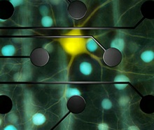
Rresearchers map functional connections between retinal neurons at single-cell resolution
08 Oct 2010
By comparing a clearly defined visual input with the electrical output of the retina, researchers at the Salk Institute for Biological Studies were able to trace for the first time the neuronal circuitry that connects individual photoreceptors with retinal ganglion cells, the neurons that carry visuals signals from the eye to the brain.
 Their measurements, published in the Oct. 7, 2010, issue of the journal Nature, not only reveal computations in a neural circuit at the elementary resolution of individual neurons but also shed light on the neural code used by the retina to relay color information to the brain.
"Nobody has ever seen the entire input-output transformation performed by complete circuits in the retina at single-cell resolution," says senior author E.J. Chichilnisky, Ph.D., an associate professor in the Systems Neurobiology Laboratories. "We think these data will allow us to more deeply understand neuronal computations in the visual system and ultimately may help us construct better retinal implants."
One of the essential elements that made the experiments possible was the unique neural recording system developed by an international team of high-energy physicists from the University of California, Santa Cruz; the AGH University of Science and Technology, Krakow, Poland; and the University of Glasgow, UK. This system is able to record simultaneously the tiny electrical signals generated by hundreds of the retinal output neurons that transmit information about the outside visual world to the brain.
These recordings are made at high-speed (over ten million samples each second) and with fine spatial detail, sufficient to detect even a locally complete population of the tiny and densely spaced output cells known as "midget" retinal ganglion cells.
neural recording system


